Is your myopia "just" a vision defect or a serious eye disease?
Myopia is the most common vision defect, and its prevalence has reached epidemic proportions. Among children alone, vision defects are diagnosed in half of all young patients, with myopia accounting for over 60% of cases, astigmatism for over 30%, and hyperopia for over 15%. Failure to diagnose a vision defect before the age of 10–12 usually results in amblyopia (lazy eye). Amblyopia is a condition where blurred vision deprives the eye of the essential stimulus needed to develop sharp sight (deprivation amblyopia). A significant correlation has been observed between the development of myopia and one’s profession, alongside the protective role of sunlight exposure. Myopia develops much less frequently in individuals whose natural working environment involves distance vision and natural light (such as sailors or pilots). Conversely, the risk is significantly higher for those working at close range under artificial lighting.
A significant risk factor for the progression of myopia in young individuals is the excessive strain placed on the lens and the ciliary muscle during near-vision tasks—the two structures that form the accommodation mechanism. Prolonged work at close range leads to chronic muscle spasm and lens bulging, which provides the additional optical power necessary for clear near vision. This phenomenon of sustained near-point visual strain is the most critical modifiable factor in the development of myopia, especially in younger patients. By maintaining proper visual hygiene, it is possible to reduce the additional risk of myopia progression caused by prolonged near-range work and the phenomenon described above. Regular breaks during close-up tasks are essential. It is highly recommended to follow the 20-20-20 rule: every 20 minutes of near-vision work, shift your gaze for at least 20 seconds to an object located at least 6 meters (20 feet) away.
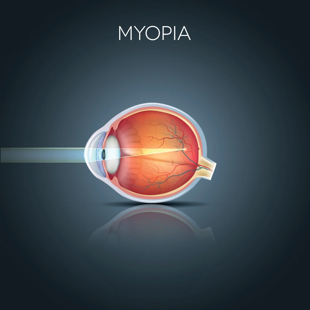
In the case of myopia, the patient’s eyeball is too long relative to the focusing light rays. As a result, the image of objects is formed in front of the retina instead of directly on it. Consequently, distant objects appear blurry—the blurriness increases with the distance from the object and the severity of the myopia. Objects seen at close range, however, remain clear. An excessive eyeball length is the most common cause of shortsightedness, known as axial myopia. In the case of refractive myopia—a less common cause—light rays are refracted too sharply due to an irregular structure of the cornea or lens, while the eyeball length itself remains normal.
Besides physiological myopia, which is by far the most common form of this condition, there is also the rare pathological myopia (also referred to as degenerative or malignant myopia). In this form, a high degree of shortsightedness is the patient’s least concern, as pathological myopia is a disease, not just a vision defect. It involves the development of degenerative changes (tissue damage) across all layers of the eye wall: the sclera, the choroid, and the innermost and most delicate layer—the retina.
Laser vision correction is contraindicated in this form of myopia. During the qualification visit, we place particular emphasis on a thorough examination of the retinal periphery and the posterior pole to detect any signs of this specific type of myopia.
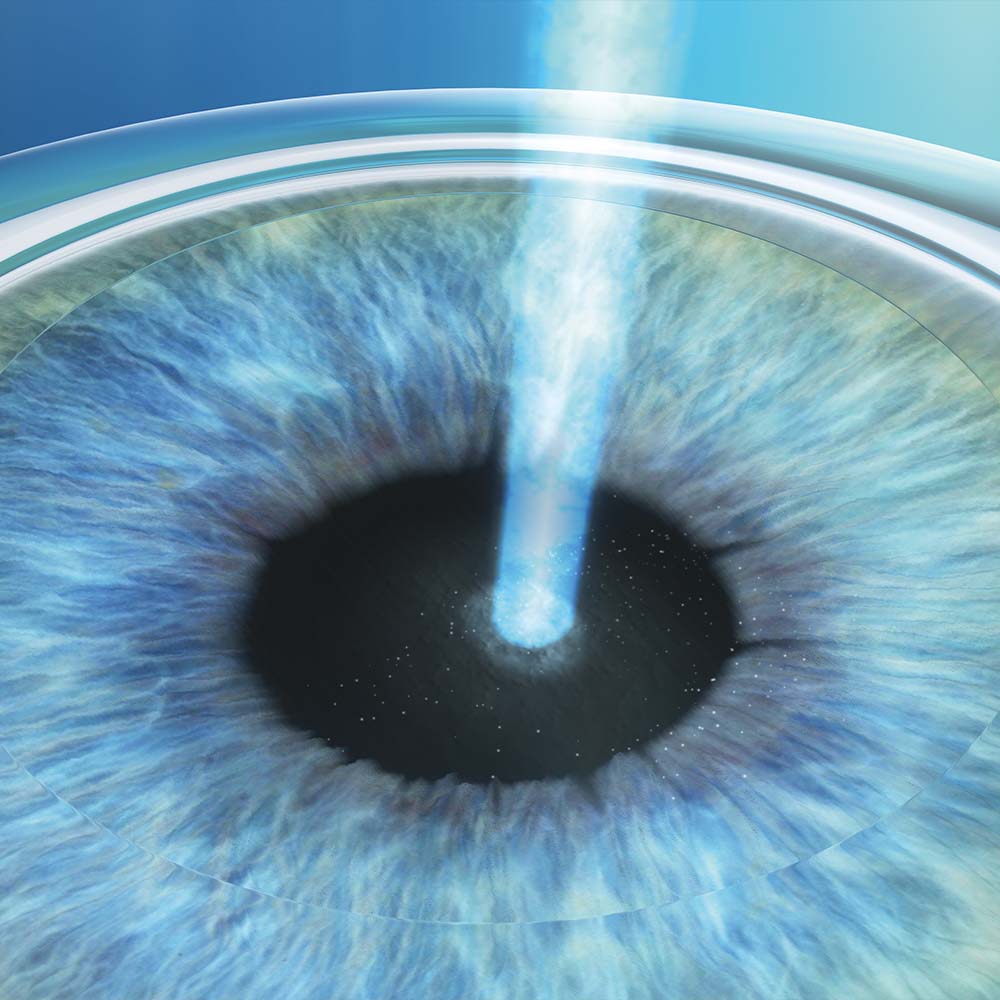
Laser vision correction for myopia involves reshaping the central part of the cornea to reduce its curvature. This allows for an extremely precise reduction in the cornea’s optical power, ensuring that images are formed directly on the retina rather than in front of it. As a result, the patient’s eyes regain optimal visual acuity.
How is myopia treated?
SMILE (myopia, myopic astigmatism)
FemtoLasik (myopia, myopic astigmatism)
EBK, PRK (low myopia with potential low astigmatism)
Phakic IOLs high myopia, high myopic astigmatism
Vision Correction Methods for Myopia
ReLex Smile
Who is ReLex SMILE for?
Patients with shortsightedness (myopia) up to -10.0 diopters.
Patients with myopic astigmatism up to -5.0 diopters, with an accompanying spherical error up to -10.0 diopters.
Why choose ReLex SMILE?
Minimal corneal interference: Uses a micro-incision (microport) only 2.8 millimeters long.
Minimal disruption to the corneal nerve plexus.
Maximum corneal stability and strength following the procedure.
Lowest susceptibility to the effects of potential future eye injuries.
One of the specialized methods for treating selected cases of keratoconus (Aztec protocol).
Shortsightedness (Myopia), High Shortsightedness (High Myopia), Myopic Astigmatism, Presbyopia in Myopia
Patients with farsightedness (hyperopia) up to +6.0 diopters.
Patients with astigmatism up to 6.0 diopters, with an accompanying spherical error up to -10.0 diopters.
Patients with near vision impairment (presbyopia).
The procedure can also be used for shortsightedness (minus errors), although this is less common due to the availability of the SMILE technology alternative.
Why choose FemtoLasik?
High EMO (Effective Modifying Power) coefficient: This method is highly effective for treating vision impairments that are more resistant to correction, such as farsightedness (hyperopia), hyperopic astigmatism, and complex astigmatism.
Easy access for future adjustments: Due to the ease of accessing the treatment zone (laser area), this method is an excellent choice for patients who may require re-correction in the future (those with farsightedness developing after age 40 or presbyopia)
Combined correction protocols: FemtoLasik allows for the simultaneous application of protocols designed to correct both distance and near vision, such as Presbyond and Presbymax.
Patients with shortsightedness (myopia) up to -3.5 diopters, possibly with accompanying astigmatism.
Patients with farsightedness (hyperopia) up to +2.5 diopters.
Patients with astigmatism up to ±2.5 diopters.
Why choose Trans-PRK?
Trans-PRK is a surface procedure in which the corneal epithelium is removed non-contact using an excimer laser. The acronym Trans-PRK stands for transepithelial PRK.
Using a laser for epithelial removal ensures that the stroma is free from minor irregularities before the corrective procedure. The stromal bed is perfectly even before the vision correction process begins, maintaining uniform humidity and temperature conditions across its entire surface.
Since there is no need to create a corneal flap or use the epithelium as a “masking substance,” this method can be carefully applied to corneas with minor irregularities, as well as in selected cases of keratoconus.
Mild Nearsightedness (Mild Myopia) and Myopic Astigmatism, Mild Farsightedness (Mild Hyperopia) and Hyoperopic Astigmatism, Mild Astigmatism
Patients with shortsightedness (myopia) up to -3.5 diopters, including those with accompanying astigmatism.
Patients with farsightedness (hyperopia) up to +2.5 diopters.
Patients with astigmatism up to ±2.5 diopters
Why choose EBK?
EBK is a surface procedure where the corneal epithelium is removed mechanically—without the use of alcohol.
The absence of alcohol in this method ensures a faster regrowth of the corneal epithelium, making the post-operative recovery period significantly milder and more comfortable.
Since there is no need to create a corneal flap, EBK is an excellent option for patients with thin corneas
Mild Shortsightedness (Mild Myopia) and Myopic Astigmatism, Mild Farsightedness (Mild Hyperopia) and Hyperopic Astigmatism, Mild Astigmatism
Phakic Lens Implantation (pIOL) Phakic lens implantation is performed to correct vision in patients for whom laser vision correction is either contraindicated or would not offer the expected results. The procedure involves implanting an artificial lens made of a specialized composite material called Collamer. Collamer is a combination of naturally occurring collagen, which provides high biocompatibility while maintaining durability and transparency. Phakic lenses are implanted adjacent to the patient’s natural lens. Preserving the eye’s own lens is a key advantage, as the patient’s accommodation remains unchanged after the procedure.
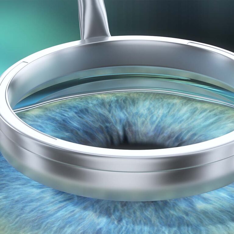
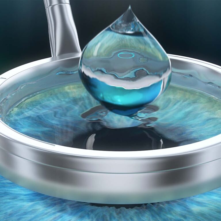
The Procedure The implantation is performed under local anesthesia after pupil dilation and takes approximately 30 minutes. The first step involves creating a corneal micro-incision (microport) 2–3 mm in length. A sterile gel (viscoelastic) is then injected into the eye chamber, allowing for the implantation of a folded lens without touching the eye’s internal structures—including the patient’s own lens and cornea. Once implanted, the lens begins to unfold spontaneously due to body temperature and the presence of the viscoelastic. After the lens is fully unfolded, the surgeon positions the phakic lens correctly in the posterior chamber. The corneal micro-incision seals itself and does not require sutures.
Reversibility Phakic lens implantation is a reversible procedure. In the event that a patient develops cataracts later in life, the clouded natural lens is removed along with the phakic lens. A standard intraocular lens (IOL) is then implanted to compensate for the power of both previous lenses, ensuring the patient’s visual outcome remains consistent.
High Shortsightedness (High Myopia), High Farsightedness (High Hyperopia), High Astigmatism
I had the SMILE procedure and I'm delighted. The doctor is a true professional – everything was thoroughly explained to me before the surgery, and I felt safe and at ease during the procedure. The procedure itself was quick and painless, and the results exceeded my expectations – my vision is excellent, better than ever before. Post-operative care was also top-notch; I could always count on answers to my questions and support.
I wholeheartedly recommend him to anyone considering vision correction – it was one of the best decisions of my life!
I highly recommend Dr. Marcin as a top eye surgery specialist in Warsaw. The procedure was totally painless and the service l have gotten was great. Now, l can see this World properly thanks to the Doctor. My sincere recommendation to everyone.
I had a lot of concerns about vision correction. I was afraid something would go wrong and I'd have to deal with the consequences. Ultimately, I went for a consultation with Dr. Smorawski on a recommendation, as he had performed the procedure on a family member a few years ago. And that was the moment I believed him. Dr. Smorawski is full of empathy and patience, personally greets and gets to know each patient, answers all questions honestly, is extremely knowledgeable and professional, and his personalized approach makes you feel completely cared for. This isn't a chain store where you have to wait for a hotline to be connected, so in case of an emergency, you can count on him.
Ms. Agata, who supports the doctor, also inspires great trust, offers advice, and patiently answers questions. 😉 I've been having my vision correction done for a few weeks now, from -3 to 0, and my quality of life has improved significantly. I can only say that I regret waiting so long. I would choose the same place again, and only Dr. Smorawski. I recommend it 100%.
If you are looking for an ophthalmologist I highly recommend the Smorawski Okulistyka clinic.
Dr. Marcin Smorawski performed my SMILE procedure lately, and I had the pleasure of receiving his excellent care. From the very first consultation, Dr. Smorawski impressed me with his professionalism. He patiently answered all of my questions and took the time to thoroughly explain every step of the process. His ability to make complicated medical concepts understandable gave me the confidence to feel well-informed and secure in my choice. The results of the LASIK surgery have been life-changing, and I owe it all to Dr. Smorawski’s expertise.
Myopia - consultation with a specialist
Are you struggling with myopia and want to effectively get rid of this vision defect? Contact us to determine the treatment method that is best for your case.
To provide the best experiences, we use technologies like cookies to store and/or access device information. Consenting to these technologies will allow us to process data such as browsing behavior or unique IDs on this site. Not consenting or withdrawing consent, may adversely affect certain features and functions.
Functional
Always active
The technical storage or access is strictly necessary for the legitimate purpose of enabling the use of a specific service explicitly requested by the subscriber or user, or for the sole purpose of carrying out the transmission of a communication over an electronic communications network.
Preferences
The technical storage or access is necessary for the legitimate purpose of storing preferences that are not requested by the subscriber or user.
Statistics
The technical storage or access that is used exclusively for statistical purposes.The technical storage or access that is used exclusively for anonymous statistical purposes. Without a subpoena, voluntary compliance on the part of your Internet Service Provider, or additional records from a third party, information stored or retrieved for this purpose alone cannot usually be used to identify you.
Marketing
The technical storage or access is required to create user profiles to send advertising, or to track the user on a website or across several websites for similar marketing purposes.
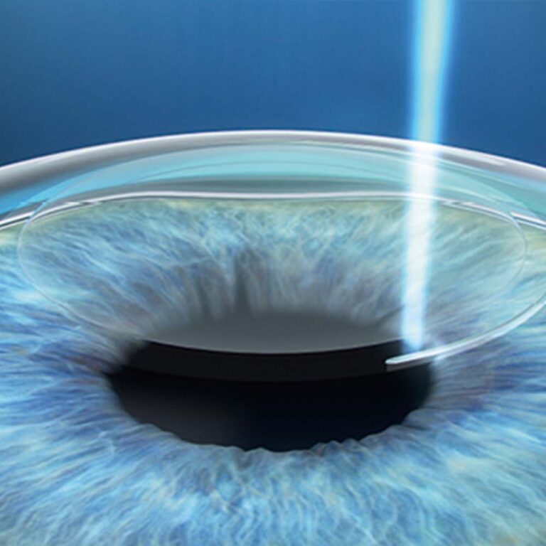
The corneal surface (the outer part) is its most valuable layer. SMILE technology leaves this vital outer section intact, as the procedure removes tissue only from the deeper layers of the cornea. As a result, corneal stability and strength after the procedure are higher than with any other method. For example, correcting 4 diopters of shortsightedness using the SMILE method leaves the cornea significantly more resilient compared to other correction techniques such as Femto-LASIK or PRK.
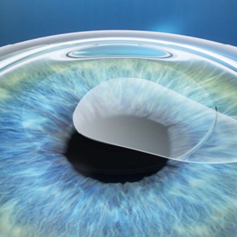
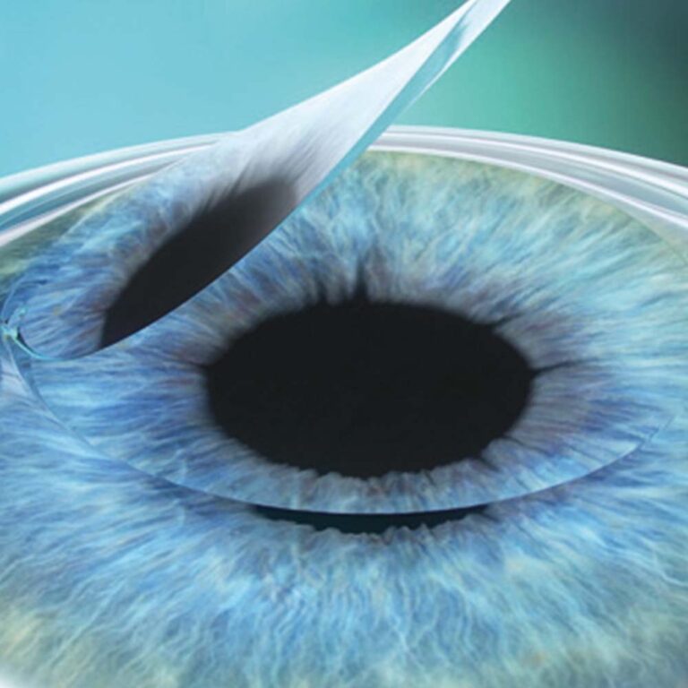
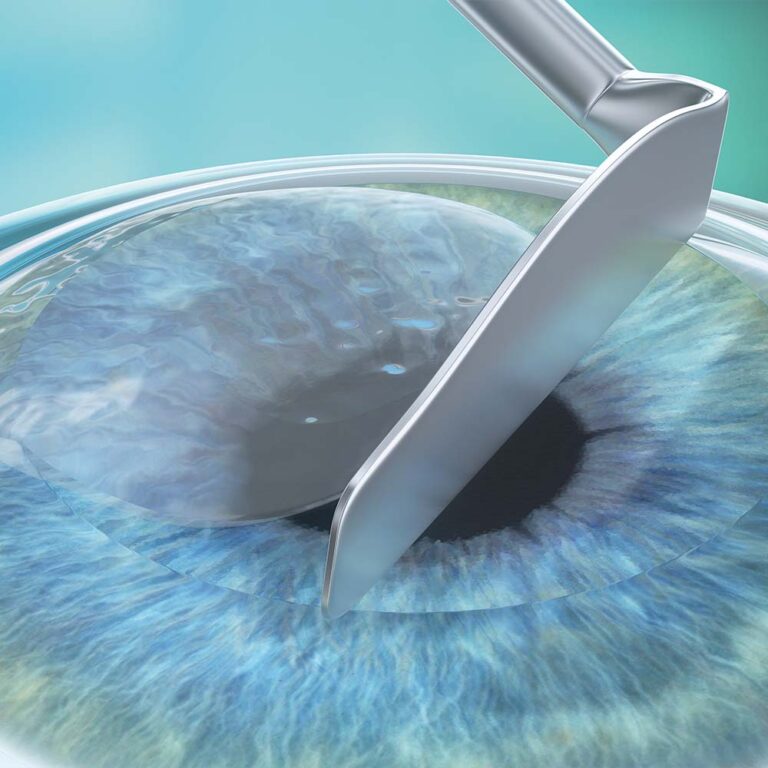
SMILE technology works by creating a thin disc of tissue (lenticule) within the deeper layers of the cornea. Once this disc is removed through a tiny micro-incision, the cornea flattens and its refractive power is reduced. This effectively eliminates shortsightedness or myopic astigmatism—the two primary vision impairments for which SMILE technology is utilized. Consequently, the procedure does not require a corneal flap.
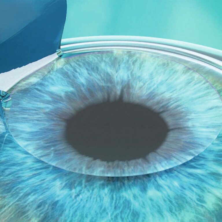
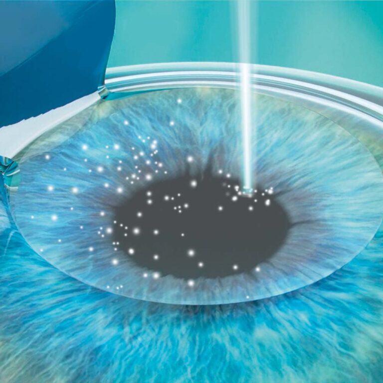
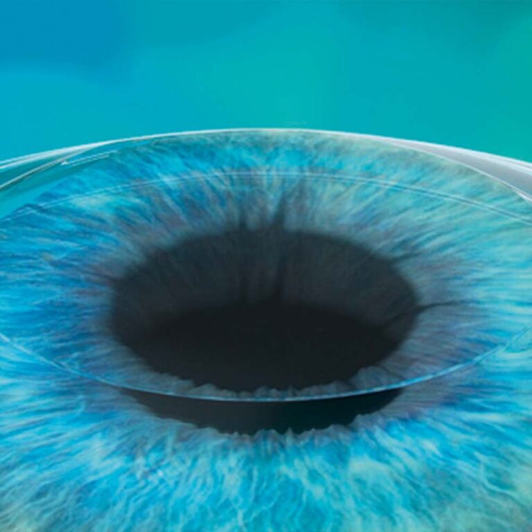
Hyperopia, hyperopic astigmatism, and astigmatism are the most resistant refractive errors to correct. To eliminate farsightedness, a change is required that results in increased corneal steepening. Steepening the corneal dome is significantly more challenging to achieve with laser correction than flattening it. This creates the need for a procedure with a higher EMO (Effective Modifying Power), which is more effective at reshaping the tissue. Femto-LASIK is such a procedure. It is a two-stage treatment. The first stage, which involves creating a corneal flap, simultaneously reduces the rigidity (softens) of the cornea. Once prepared this way, the cornea becomes much more pliable and susceptible to reshaping during the second stage. In the second stage, an excimer laser is applied to the corneal stroma exposed under the flap. In the treatment of presbyopia, we take advantage of the practicality and easy access provided by the presence of the corneal flap. As we age, the demand for visual power for near-distance work increases. To meet this growing need, we can compensate for the increasing near-vision impairment through a minimally invasive and simple method: a flap lift.
Why is Trans-PRK correction more predictable?
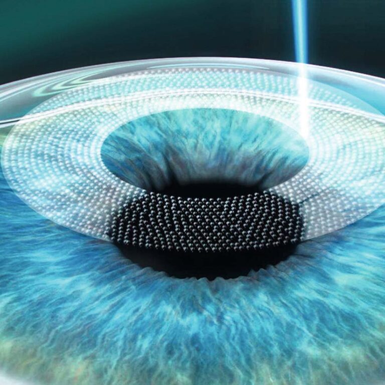
The epithelium covers the corneal stroma and is its outermost layer. In surface methods (EBK, Trans-PRK, Topo-guided-PRK), the first step is the removal of the corneal epithelium. Due to the specific way the laser operates, removing the epithelium with a laser leaves the cornea highly uniform in terms of surface texture and hydration levels. The correction stage follows immediately after the epithelial removal phase, making the overall result significantly more predictable compared to manual removal.
What does it mean that the epithelium can be used as a masking substance?
The human eye is not perfect; minor irregularities often occur on the corneal surface. These are frequently compensated for by the overlying epithelium. Thus, the epithelium acts as a tissue that masks imperfections in the corneal stroma. When removing the epithelium manually, these surface imperfections are exposed rather than eliminated. However, by using an excimer laser for epithelial removal, we eliminate corneal irregularities along with the epithelium, achieving an optically ideal surface.
EBK and PRK are surface vision correction methods in which the first, preparatory stage is the removal of the corneal epithelium. In PRK, the epithelium is removed manually using a special solution that loosens the connection between the epithelium and the corneal stroma. In EBK, also a manual method, a specialized device called an epikeratome is used to remove the epithelium. The corneal epithelium regenerates—or regrows—within several dozen hours, forming a perfectly smooth and even surface. The epithelium has a massive impact on visual quality, and even microscopic irregularities on its surface can drastically impair vision.
The epithelium is a fascinating structure with the ability to mask any imperfections of the underlying stroma. This characteristic is very often utilized in the treatment of corneal irregularities and keratoconus.
Phakic lenses offer the widest range of vision correction of all available methods. It is important to emphasize that they serve as both an alternative to laser vision correction and a viable treatment option for patients where laser treatment would not deliver satisfactory results. With this correction method, there is no tendency for regression (recurrence of the refractive error).


 4.8Based on 38 reviewspowered by Google
 aa13:26 02 Apr 26I recommend it 100%. Thanks to specialists like you, I regain my faith in the medical profession. 10/10. KK
 Bartosz Bober11:48 24 Sep 25I had the SMILE procedure and I'm delighted. The doctor is a true professional – everything was thoroughly explained to me before the surgery, and I felt safe and at ease during the procedure. The procedure itself was quick and painless, and the results exceeded my expectations – my vision is excellent, better than ever before. Post-operative care was also top-notch; I could always count on answers to my questions and support.
 Renata19:12 10 Jul 25Very good doctor. Friendly staff. Thanks to the correction, I have a new life. I highly recommend it.
 Olena Shmanko12:52 06 Mar 25I highly recommend Dr. Marcin as a top eye surgery specialist in Warsaw.
 Monika K08:53 23 Dec 24I had a lot of concerns about vision correction. I was afraid something would go wrong and I'd have to deal with the consequences. Ultimately, I went for a consultation with Dr. Smorawski on a recommendation, as he had performed the procedure on a family member a few years ago. And that was the moment I believed him. Dr. Smorawski is full of empathy and patience, personally greets and gets to know each patient, answers all questions honestly, is extremely knowledgeable and professional, and his personalized approach makes you feel completely cared for. This isn't a chain store where you have to wait for a hotline to be connected, so in case of an emergency, you can count on him.
 Catherine Jofi09:43 20 Aug 24If you are looking for an ophthalmologist I highly recommend the Smorawski Okulistyka clinic.